BMD clinical trials: DXA calibration pitfalls
A bone mineral density trial can lose analytical validity before the first biological endpoint is analyzed. The failure point is often not the intervention. It is the DXA measurement chain.
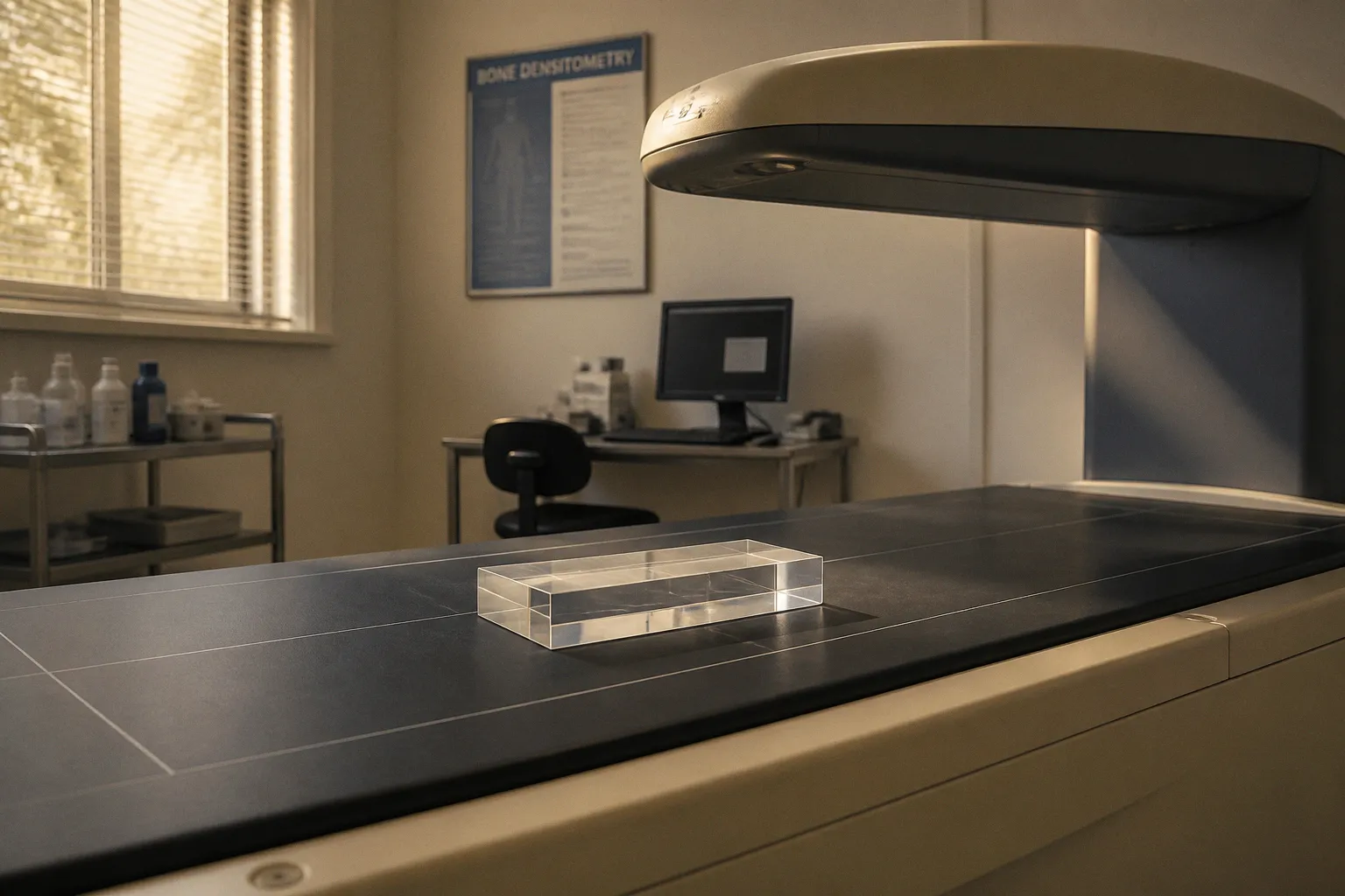
Comparative testing has shown that Hologic DXA systems can report lumbar spine BMD values 14% to 20% lower than GE Lunar systems. At femoral sites, the difference can reach 8% to 17%. These are not random fluctuations. They are systematic offsets associated with scanner manufacturer, hardware configuration, software, acquisition protocol, and operator execution.
For vitamin D bone density trials, this problem is direct. The endpoint is commonly a change in BMD measured by dual-energy X-ray absorptiometry. If the expected biological change is small, an uncorrected scanner offset can exceed the treatment signal. The resulting dataset may show a false treatment effect, suppress a real effect, or increase variance until the study becomes statistically inefficient.
The systematic bias of a multi-manufacturer DXA environment
DXA does not measure mineral mass by direct chemical extraction. It estimates areal bone mineral density from two X-ray energy levels and the attenuation behavior of bone and surrounding tissue. The final value depends on the scanner’s detector system, calibration model, edge-detection algorithm, reference database, software version, and analysis procedure.
That makes BMD a platform-dependent measurement.
The reported value is expressed in g/cm², but the unit does not guarantee interchangeability. A lumbar spine measurement from one manufacturer is not automatically equivalent to a measurement from another manufacturer simply because both instruments are labeled DXA and both report g/cm².
In a single-center trial using one stable scanner, the main concern is measurement precision. In a multicenter trial, the problem expands. The study must control both random error and systematic bias.
Random error affects repeatability. It is visible as dispersion around a mean value. Systematic bias shifts the entire measurement distribution. It can remain hidden when each site performs well internally but uses a different scanner platform.
This distinction determines the calibration strategy:
- Precision control asks whether the same patient can be measured consistently on the same system.
- Cross-calibration asks whether measurements from different systems can be placed on a common scale.
- Quality control verifies that scanner performance remains stable over time.
- Data validation determines whether observed differences represent biological change or instrumentation effects.
A trial that establishes only local repeatability has not solved the multicenter problem. It has measured the consistency of separate local systems.
A low coefficient of variation does not make two scanners equivalent. It only shows that each scanner may be internally repeatable.
The risk is greatest when the study randomizes participants across sites without balancing scanner type by treatment arm. Suppose one treatment group is overrepresented at sites using a platform with systematically higher spine BMD values. The baseline distribution may be corrected statistically, but the longitudinal change remains vulnerable if follow-up scans occur on different platforms or if hardware is replaced during the study.
The issue is also relevant to vitamin D research because expected skeletal effects may be gradual. Vitamin D status, serum 25-hydroxyvitamin D concentration, calcium absorption kinetics, and bone remodeling do not necessarily produce large short-term changes in areal BMD. The smaller the biological signal, the greater the relative influence of the measurement system.
Quantifying the Hologic–GE Lunar discrepancy
The reported manufacturer differences are large enough to invalidate direct pooling of raw values.
Comparative testing identified the following offsets:
| Measurement region | Hologic system compared with GE Lunar system | Analytical implication |
|---|---|---|
| Lumbar spine, L1–L4 | 14% to 20% lower BMD readings | Large platform effect; raw values should not be pooled |
| Femoral sites | 8% to 17% lower BMD readings | Site-specific bias remains clinically relevant |
| GE Lunar iDXA versus GE Lunar Prodigy, lumbar spine | 1.54% higher on iDXA | Same-manufacturer upgrades can create measurable shifts |
| GE Lunar iDXA versus Prodigy, dual femur | 1.28% to 1.56% higher on iDXA | Hardware generation affects longitudinal comparability |
The first comparison is not a minor correction factor. A 14% to 20% difference at the lumbar spine can be many times larger than the least significant change expected from a well-controlled individual measurement. Treating those values as interchangeable introduces a platform effect into the endpoint.
A trial database may therefore contain two different types of signal:
1. A true difference caused by treatment, disease progression, or nutritional intervention.
2. A measurement difference caused by the scanner platform or transition between platforms.
The database does not identify the source automatically. That separation must be designed before acquisition.
Why manufacturer labels are insufficient
The phrase “Hologic versus GE Lunar” is useful for identifying a broad source of bias, but it is not a complete calibration model. Systems within a manufacturer family may differ by hardware generation, detector configuration, software, reference database, and analysis behavior.
The GE Lunar iDXA and GE Lunar Prodigy example demonstrates this point. The iDXA reported lumbar spine BMD 1.54% higher than the Prodigy. For dual femur measurements, the increase ranged from 1.28% to 1.56%.
Those offsets are smaller than the reported Hologic–GE Lunar differences, but they are still operationally significant. In a longitudinal trial, the hardware change may occur between scheduled visits. A patient scanned on the Prodigy at baseline and on the iDXA at follow-up can appear to gain BMD even if the underlying skeletal status is unchanged.
The direction of the error also matters. A fixed offset between two systems does not behave like random noise. It can produce an apparent step change at the time of the instrument transition.
Site-specific behavior must remain visible
A lumbar spine calibration equation should not be assumed to correct femoral sites. DXA algorithms interact with anatomical geometry, soft tissue composition, positioning, and region-of-interest placement. The discrepancy observed at L1–L4 is not a universal proxy for the proximal femur.
This is one reason a multicenter protocol should preserve the original scanner identity, model, software version, acquisition date, analysis site, and technologist identity for every scan. A final BMD value without its measurement metadata is an incomplete analytical record.
For vitamin D bone density trials, the minimum useful metadata set includes:
- Scanner manufacturer and model.
- Software version and hardware generation.
- Site identifier.
- Scan date and visit number.
- Technologist identifier.
- Anatomical region and vertebral levels analyzed.
- Whether the scan was acquired before or after a scanner service, upgrade, or replacement.
- Whether the analysis was performed locally or centrally.
- Any reanalysis or region-of-interest adjustment.
These fields do not correct an offset. They make the offset detectable and allow the statistical analysis to account for it.
Intra-manufacturer variability: the hidden cost of hardware upgrades
Replacing a scanner from the same manufacturer is often treated as a routine operational event. In a clinical trial, it is a measurement-system change.
The assumption that a newer model is automatically compatible with the previous model is not valid. Improved detector performance, altered calibration coefficients, revised software, and different edge-detection behavior can modify the reported BMD value.
The GE Lunar iDXA and Prodigy comparison provides a practical example. The iDXA produced higher readings at the lumbar spine and dual femur despite both systems belonging to the same manufacturer family. The difference was systematic. The hardware upgrade therefore introduced a potential discontinuity into any longitudinal series.
A scanner replacement should be handled as a protocol event, not as ordinary maintenance. The trial team must determine:
1. Whether the old and new systems are both available for a defined overlap period.
2. Whether the same patient can be scanned on both systems under controlled conditions.
3. Whether the study requires local in vivo cross-calibration.
4. Whether the site’s technologists have established precision on the new system.
5. Whether the central analysis pipeline can preserve and apply the resulting equations.
6. Whether scans obtained before the transition require reanalysis.
7. Whether the transition date must be included as a covariate or stratification variable.
Phantom measurements are useful for monitoring scanner stability. They do not, by themselves, establish that patient measurements are interchangeable across platforms. A phantom has a fixed composition and geometry. Human measurements include positioning variability, soft tissue effects, anatomical structure, and region-of-interest decisions.
Phantom stability confirms instrument behavior on the phantom. It does not prove equivalence of in vivo patient measurements after a hardware transition.
The distinction is operationally important. A scanner may pass routine phantom quality control while still requiring a cross-calibration equation for patient-level data. These are different control layers.
Software changes are also part of the measurement system
A hardware upgrade is obvious. A software update can be less visible and equally disruptive. Changes in segmentation algorithms, reference databases, default analysis settings, or automated edge detection may alter the reported value without changing the X-ray source or detector.
The trial master file should therefore record software changes with the same discipline applied to hardware changes. The relevant question is not whether the update was described as minor. The relevant question is whether it modified the measurement or analysis pathway.
If the answer is uncertain, the transition should be treated as a potential source of systematic error until evaluated.
Establishing precision: CV and least significant change
Cross-calibration cannot compensate for poor local precision. Each site must first demonstrate that its operators and scanner can produce repeatable measurements.
Short-term human precision trials have reported coefficient of variation values from 0.7% to 2.2% in the lumbar spine and proximal femur. Reported least significant change values ranged from 0.018 to 0.048 g/cm².
The coefficient of variation describes relative measurement variability. The least significant change translates precision into an absolute threshold. It defines the minimum observed change that can be interpreted as exceeding expected measurement error under the specified confidence convention.
These values are not universal constants. They depend on the scanner, site, technologist, anatomical region, patient population, positioning procedure, analysis method, and precision study design.
A site should not import an LSC from a publication and apply it without local qualification. The published range establishes the scale of the problem. It does not replace a site-specific precision assessment.
Why LSC matters in a vitamin D trial
Vitamin D trials often evaluate changes in bone-related outcomes alongside serum 25-hydroxyvitamin D. A biochemical increase in vitamin D status can be readily observed, while the skeletal response may be smaller, slower, or dependent on baseline deficiency and calcium availability.
If the measured BMD change is below the site’s LSC, the result cannot be confidently separated from measurement variability at the individual level. At the trial level, repeated measurements and statistical modeling can improve estimation, but they cannot eliminate systematic offsets between scanners.
The practical interpretation is conditional:
- A change smaller than the local LSC is not automatically evidence of no biological effect.
- A change larger than the LSC is not automatically evidence of treatment efficacy if the scanner changed.
- A between-group difference is not secure if treatment allocation is confounded with scanner platform.
- A statistically significant result can still be analytically compromised by unmodeled instrument effects.
Precision testing must include the operator
The technologist is part of the measurement system. Patient positioning, scan acquisition, vertebral identification, femoral rotation, region-of-interest placement, artifact handling, and analysis decisions all contribute to variability.
A site that changes operators during a trial may experience a precision shift even when the scanner remains unchanged. Training records and operator-specific precision data are therefore relevant to endpoint validation.
The same applies to central reanalysis. Central reading can reduce certain forms of local analytical variability, but it does not erase acquisition differences caused by positioning or scanner physics. The acquisition and analysis stages must be treated separately.
Standardizing multicenter data beyond phantom calibration
A valid multicenter DXA program requires a layered control system. No single procedure addresses all sources of error.
The sequence should begin before the first participant is scanned.
1. Define the platform architecture
The protocol should specify which scanner families are acceptable, which models require separate calibration strata, and whether new sites can be added after study initiation. A broad statement permitting any DXA system creates avoidable heterogeneity.
The study should also define the primary BMD regions. Lumbar spine and proximal femur are not interchangeable endpoints. Each region requires its own precision and cross-calibration logic.
2. Establish baseline quality control
Every participating site should document routine quality control procedures before patient enrollment. This includes phantom monitoring, service records, scanner status, software version, and technologist qualification.
The purpose is not to create administrative volume. It is to establish the pre-intervention state of the measurement system.
3. Perform local precision assessment
Precision should be quantified for the operators who will acquire trial scans. The assessment should reflect the study’s target regions and acquisition procedure.
The resulting CV and LSC should be retained as site-level analytical parameters. If a site has materially weaker precision than the rest of the network, the response should be technical remediation, additional training, or exclusion from a specific endpoint—not silent pooling.
4. Conduct cross-calibration when platforms differ
Cross-calibration requires more than comparing manufacturer specifications. The trial must establish how values from one platform relate to values from another for the relevant anatomical regions.
The exact universal mathematical formula for every legacy DXA model and current platform does not exist. Local in vivo testing may be required, particularly when a hardware change occurs during an active longitudinal study.
The calibration equation must be region-specific and documented. It should not be applied outside the population, scanner pair, anatomical site, or acquisition conditions for which it was derived without justification.
5. Control scanner transitions
A replacement or upgrade should trigger a predefined transition procedure. The protocol should identify whether patients are rescanned on both systems, whether a bridging subset is used, and how the transition is represented in the analysis dataset.
If an old scanner is removed before bridging is completed, the opportunity to characterize the discontinuity may be lost. Retrospective statistical correction is then less secure because the relationship between systems has not been measured under trial conditions.
6. Preserve untransformed and transformed values
The raw instrument output should be retained. If cross-calibration is applied, the transformed value should be stored as a separate variable with the equation version, calibration dataset, region, and implementation date.
Overwriting the original value prevents auditability. It also makes later sensitivity analyses difficult.
A robust database should distinguish at least:
- Original scanner-reported BMD.
- Cross-calibrated BMD.
- Scanner and model.
- Calibration stratum.
- Transition status.
- Site-specific precision parameters.
- Analysis version.
- Flag for artifact, reanalysis, or protocol deviation.
7. Predefine statistical handling
The statistical analysis plan should state how scanner platform, site, hardware transition, and calibration status enter the model. This is particularly important when randomization is not balanced across scanners.
Possible approaches may include stratification, covariate adjustment, mixed-effects modeling, or sensitivity analyses by scanner family. The appropriate method depends on the study design and the calibration data. No generic correction should be presented as universally sufficient.
The data should also be reviewed for discontinuities at scanner transition dates. A sudden shift in mean BMD coincident with an upgrade is an analytical signal. It should not be dismissed as biological variation without examination.
What a validated BMD dataset should demonstrate
A defensible BMD dataset should allow an independent reviewer to answer five questions.
Was the scanner stable?
Routine phantom quality control and service documentation should show whether the instrument remained within its expected operating range. A drift pattern may indicate hardware or calibration deterioration.
Was the operator precise?
The site should have evidence of technologist-level repeatability for the relevant anatomical regions. General certification is not equivalent to study-specific precision.
Were platforms comparable?
If multiple manufacturers or hardware generations were used, the study should document the cross-calibration method. Raw values should not be treated as directly comparable solely because they share the same unit.
Were transitions controlled?
Any scanner replacement, software update, service intervention, or operator change should have a date and an associated assessment. The absence of a transition record is itself a data-quality limitation.
Can the reported change exceed the measurement system?
The observed treatment difference should be interpreted against the precision profile, including local LSC and cross-platform uncertainty. A numerical change without this context is incomplete.
The International Society for Clinical Densitometry provides official positions addressing cross-calibration, least significant change, and quality assurance in clinical environments using multiple scanners. Those positions should be incorporated into protocol development rather than consulted only after inconsistent results appear.
DXA has been used for bone mass measurement under FDA approval since 1988. Its long clinical history does not remove the need for calibration discipline. Mature technology still produces invalid comparisons when measurement conditions change without documentation.
Common failure modes in vitamin D bone density trials
Several errors recur because they appear operationally reasonable.
Pooling manufacturer outputs by nominal unit
The mistake is simple: all instruments report g/cm², so all values are placed in one column and analyzed together. The unit standardizes the expression, not the underlying measurement response.
Using one correction factor for every skeletal site
A lumbar spine equation is applied to femoral neck, total hip, or dual femur values. This assumes that the platform difference is anatomically uniform. It is not justified without validation.
Treating same-manufacturer upgrades as neutral
The iDXA–Prodigy comparison shows that a same-manufacturer transition can produce a systematic offset. Brand continuity is not calibration continuity.
Relying on phantom data alone
A phantom can verify stability of a scanner under defined conditions. It cannot establish in vivo equivalence after a platform change. Patient positioning and soft tissue composition remain relevant variables.
Applying an external LSC without local precision testing
Published CV and LSC ranges provide reference context. They do not characterize the exact site, operator, and scanner combination used in a particular trial.
Ignoring the timing of instrument changes
A scanner upgrade is recorded as a maintenance event, but the analysis dataset lacks the transition date. The resulting step change is then misclassified as biological response or random noise.
Replacing raw data with corrected data
Once transformed values overwrite original readings, the calibration process becomes difficult to audit. Both versions are required for traceability.
A practical control matrix
The following structure separates the main control objectives:
| Control layer | Primary question | Required evidence | Failure consequence |
|---|---|---|---|
| Phantom quality control | Is the scanner operating consistently? | Longitudinal phantom records and service logs | Drift may be mistaken for patient-level change |
| Technologist precision | Can the operator reproduce the measurement? | Site- and operator-specific CV and LSC | Increased random error |
| Cross-calibration | Can platforms be placed on a common scale? | Region-specific equations or validated bridging data | Systematic inter-machine bias |
| Transition management | What changed and when? | Hardware, software, and operator change records | Unrecognized discontinuity |
| Central data review | Are values and metadata coherent? | Audit trail, reanalysis records, discrepancy resolution | Undetected protocol and analysis errors |
| Statistical adjustment | Is platform effect separated from treatment effect? | Prespecified model and sensitivity analysis | Confounded efficacy estimate |
This matrix is more useful than a single statement that the study used standardized DXA. Standardization is not a label. It is a chain of documented controls.
The technical limits of interpretation
DXA-derived BMD remains an areal measurement. It is not a complete description of bone material quality, microarchitecture, or fracture resistance. In vitamin D research, it should be interpreted alongside biochemical status, calcium intake or availability, relevant clinical outcomes, and the biological timing of remodeling.
That broader interpretation does not reduce the importance of calibration. It increases it. When BMD is one component of a multi-endpoint study, the analytical meaning of a small change depends on whether the measurement system is stable enough to support the conclusion.
The full effect of changing soft tissue composition on edge-detection algorithms across manufacturers is not completely defined. This is an additional reason to avoid overconfident universal corrections. A calibration equation validated in one setting should not be treated as a physical law for every patient, scanner, and study population.
Similarly, no single cross-calibration formula can be assumed to connect every legacy DXA model with every current platform without local evaluation. The correct response to this limitation is controlled measurement, not numerical improvisation.
Cost-benefit summary for trial design
The cost of rigorous DXA standardization is front-loaded. It includes scanner inventory, precision testing, technologist qualification, bridging scans, calibration analysis, metadata management, and predefined statistical handling.
The cost of omitting these controls appears later. It includes inflated sample-size requirements, ambiguous treatment estimates, unusable subgroup comparisons, reanalysis, protocol deviations, and potentially invalid claims about skeletal benefit.
The technical sequence is therefore strict:
1. Inventory every scanner, model, software version, and site.
2. Establish local technologist precision for each target anatomical region.
3. Define acceptable platforms and calibration strata.
4. Cross-calibrate different systems using region-specific evidence.
5. Bridge hardware transitions before relying on post-upgrade longitudinal values.
6. Retain raw, corrected, and metadata-linked measurements.
7. Compare observed changes with local LSC and cross-platform uncertainty.
8. Prespecify the statistical treatment of site and scanner effects.
A bone mineral density trial using DXA is only as comparable as its least controlled measurement transition. Manufacturer differences of 14% to 20% at the lumbar spine and 8% to 17% at femoral sites are large enough to dominate the endpoint. Same-manufacturer upgrades can also shift values, as shown by the higher readings from GE Lunar iDXA relative to Prodigy.
The benefit of calibration is not cosmetic consistency. It is the ability to distinguish vitamin D-related biological change from instrument behavior. Without that separation, the BMD result remains a measurement output, not a validated clinical conclusion.